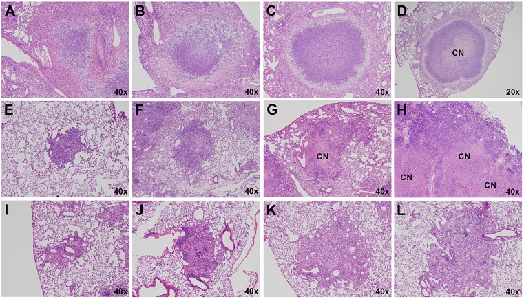
Fig. 2.
Progression of Type I, II and III lesions over time. Each panel represents a single lesion (H&E-stained) obtained from an individual representative animal (n=5) euthanized at the time point defined here. Progression of Type I lesions from individual animals at 45 (A), 50 (B), 55 (C) and 61 (D) days following aerosol infection. Progression of Type II lesions from individual animals at 20 (E), 35 (F), 38 (G) and 40 (H) days following aerosol infection. Progression of Type III lesions from individual animals at 30 (I), 35 (J), 61 (K) and 75 (L) days following aerosol infection. CN, caseous necrosis.

