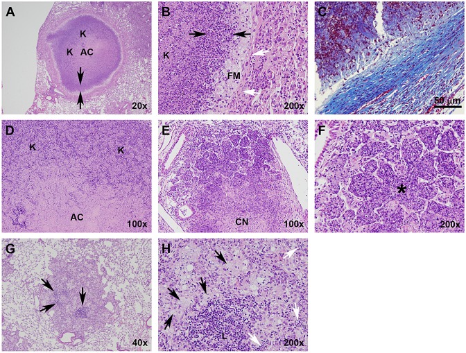
Fig. 3.
Type I, II and III lesions vary with respect to their cellular composition, organization and collagen encapsulation. (A) Type I granuloma showing the acellular caseum, distinct band of darkly stained intact neutrophils (arrows delineate margins), primarily neutrophil-derived zone of karyorrhectic debris, lightly stained foamy macrophage rim, and collagen encapsulation. (B) Higher-magnification image of A illustrating a zone of intact neutrophils (black arrows), foamy macrophage rim and collagen encapsulation (white arrows). (C) Masson's Trichrome stain for collagen along the peripheral margin of a Type I lesion. (D) Interior region of a Type I granuloma, showing the acellular caseum and karyorrhectic debris. (E) Type II lesion with a caseous necrotic interior, and extensive consolidation of peripheral alveoli. (F) Pulmonary acinus (*) completely occluded with neutrophils and cellular debris. (G) Type III lesion with distinct lymphocytic clusters (arrows). (H) Higher-magnification image of G showing foamy macrophages (black arrows) and a lymphocytic cluster (L). Small isolated pockets containing small numbers of neutrophils (white arrows) are occasionally present, especially at later time points as foamy macrophages undergo necrosis. AC, acellular caseum; K, karyorrhectic debris; FM, foamy macrophages; CN, central necrosis. H&E staining unless otherwise noted.

