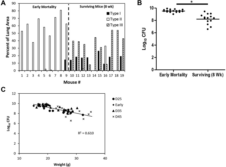
Fig. 4.
Early-mortality mice displayed primarily Type II lesions, and had higher pulmonary bacterial loads and lower body weights. (A) Early-mortality mice primarily exhibited Type II lesions, whereas the surviving mice exhibited primarily Type I and Type III lesions. Lesion area analysis was performed on H&E-stained histological sections of all five lung lobes from individual mice (n=9 for early mortality mice and n=10 for surviving mice). Early-mortality mice represent mice euthanized between day 30 and 41 post-infection owing to morbidity. Surviving mice were euthanized 8 weeks following infection. Data are expressed as the percentage of the total lung area (all five lobes) for each lesion type compared to the total lung area for individual mice. (B) Early-mortality mice (n=17) had a higher pulmonary bacterial load compared to surviving mice (n=14). Data represent the logarithms of serial dilutions of lung homogenates obtained from whole lungs of individual mice. Statistical comparison performed using Student's t-test. *P<0.0001. (C) Animal body weight was inversely correlated with pulmonary bacterial burden. Terminal body weight was taken immediately prior to euthanasia with pulmonary bacterial load expressed as log10 CFU. Early-mortality mice were euthanized between 30 and 41 days post-infection owing to morbidity, and ten mice were euthanized at 25 (D25), 35 (D35) and 45 (D45) days post-infection.

