Fig. 7.
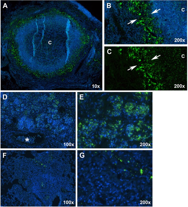
Fluorescent acid-fast staining revealed differences in bacterial numbers and spatial distribution between the three lesion types. (A) A mature Type I lesion with caseous necrosis, showing the distribution of bacteria using the SYBR Gold methodology. ‘C’, caseum. (B) Large numbers of intracellular bacilli were present as aggregates within foamy macrophages (arrows delineate margins). Intracellular bacilli were also present within intact neutrophils and large numbers of extracellular bacilli were found within the caseous necrotic, hypoxic caseum. (C) The identical image of panel B with the DAPI channel turned off to more easily visualize extracellular bacilli within the caseum. (D) A Type II lesion showing large numbers of bacteria present within the peripheral margins of the lesion and in the cellular debris within terminal bronchioles (asterisk). (E) Large numbers of bacilli were located within neutrophils that had consolidated alveoli. (F) A Type III lesion showing characteristically small numbers of primarily intracellular bacilli within a relatively large lesion. (G) Higher-magnification image showing that, within Type III lesions, bacilli occurred singly, or in relatively small aggregates, in the vicinity of much larger numbers of uninfected cells. Green, SYBR Gold-stained bacilli; blue, DAPI.

