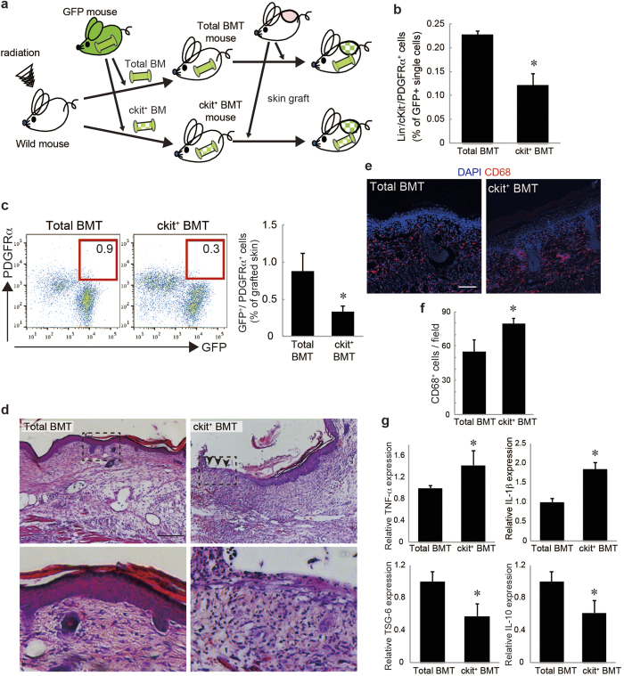
Figure 2. The reduction of endogenous PDGFRα+ mesenchymal cells delayed skin graft regeneration.
a: The skin graft model in ckit+ BMT mice. The picture was drawn by K.T. with Adobe illustrator CS6. b: The percentages of Lin−/ckit−/PDGFRα+ cells among GFP+ single cells in the BM of the ckit+ BMT mice. c: The percentages of GFP+/PDGFRα+ cells recruited into skin grafts on the total and ckit+ BMT mice. d: Skin grafts stained by H&E. The boxed regions in the upper panels (×10, bar = 200 μm) are displayed at higher magnification (×40, bar = 50 μm) in the lower panels. e: Representative images of skin grafts stained for CD68. Nuclei were stained with DAPI. Bar = 100 μm. f: The number of CD68+ cells per field was quantified. g: The expressions of TNF-α, IL-1β, TSG-6, and IL-10 in skin grafts. *P < 0.05.

