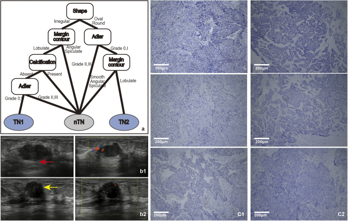
Figure 6. a.
: The model for the TN subtype followed two rules. First, the tumour shape was judged. If the shape was irregular, then the margin contour was assessed. If it was lobulate, then the calcification was judged. If calcification was absent, then the Adler degree was judged. If Adler degree was 0 or I, the tumor was classified as the TN subtype. Second, if the tumour was oval or round, then the Adler degree was judged; if the Adler was 0 or I, then the margin contour. If it was lobulate, the tumor was classified as the TN subtype. b: b1 and b2 show the ultrasound images of TN1 and TN2. From left to right, the two images show 2-dimensional and colour Doppler, respectively. The margin contour of red arrow depicts the lobulate margin of TN1. The margin contour of the yellow arrow depicts the micro-lobulate margin of TN2. c: c1 and c2 show the IHC results for the TN1 and TN2 patient (c1, ×100; c2, ×100). From top to bottom, the three images of c1 and c2 represent ER (−), PR (−), HER2 (−). Unstained tumour cells are negative (as for ER, PR and HER2 in c).

