Fig. 5.
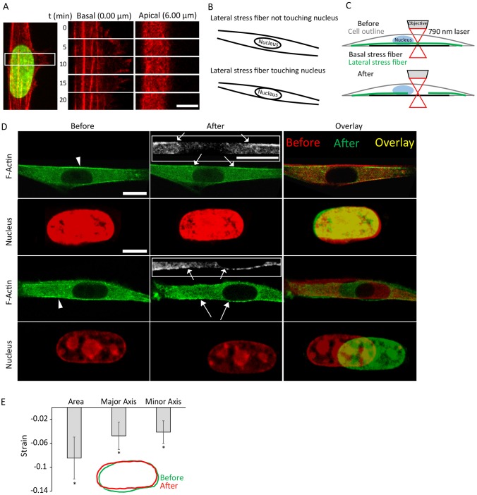
The nucleus does not expand laterally upon severing lateral stress fibers running parallel to it. (A) Kymograph during the formation and retraction of the lateral protrusion. No motion of the actin bundles underneath the nucleus is noticeable as it undergoes the local deformation. (B) Cartoon showing the two types of lateral actin stress fibers that were severed in this study. (C) A schematic showing the laser ablation experiment of lateral stress fibers. (D) Confocal images showing that laser ablation of a lateral stress fiber running parallel to the side of the nucleus either not touching it (top) or touching it (bottom) does not cause expansion in the nuclear area. Insets show the two sides of the severed stress fiber. Arrowheads indicate the severing point. Arrows indicate the two ends of the severed stress fiber. (E) Quantification of the nuclear shape change before and after lateral stress fiber severing showing a small decrease in the nuclear major and minor axes, and area upon stress fiber severing. Strains represent the change (final−initial) in the parameter divided by its initial value. Error bars represent s.e.m. N=10. *P<0.05 compared to 0. Scale bars: 6 µm (A); 10 µm (D, actin, actin inserts); 5 µm (D, nucleus).

