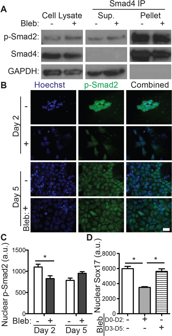
Fig. 3.

Disruption of traction forces inhibits TGF-β signaling. R1 ESCs were grown on decellularized fibroblast-derived extracellular matrix in definitive endoderm induction medium with or without 10 μM blebbistatin. (A) Blots show cells that were lysed after 2 days (left) as well as the supernatant (Sup., center) and pulldown (pellet, right) fractions of SMAD4 immunoprecipitation (IP). Blots are shown for phospho-SMAD2 (p-SMAD2), SMAD4 and GAPDH. (B) Immunofluorescent images of phospho-SMAD2 (green) and Hoechst 33342 (blue)-stained nuclei taken after 2 and 5 days are shown along with (C) quantification of the mean±s.e.m. (n>200) nuclear intensity of phospho-SMAD2. (D) Immunofluorescent images of SOX17 and Hoechst-stained nuclei were quantified (mean±s.e.m., n>1000) after 5 days of the indicated blebbistatin treatment. *P<0.05. Scale bar: 20 µm. a.u., arbitrary units.
