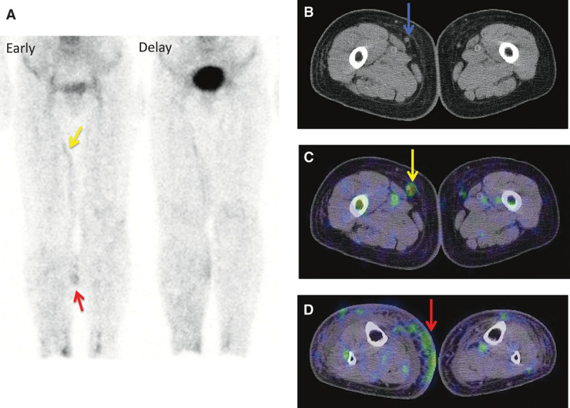
Fig. 5.

An 81-year-old woman with bilateral lower-limb lymphedema caused by a surgery for uterine cervical cancer. The right lower limb, with more severe lymphedema than the opposite side, was regarded as affected side (ISL stage: II and LEL index: 338). Both the early and delayed images show inguinal lymph node accumulation defect. Although the planar images cannot discriminate the lymphatic vessel in the medial lower thigh from dermal backflow (A, red arrow), SPECT-CT images provide the correct information as dermal backflow (D, red arrow). Furthermore, whereas the planar images cannot determine whether the tubular accumulation in the medial thigh is lymphatic vessel or vein, SPECT-CT showed vein to be the definitive answer, according to its morphological information. Therefore, this case was categorized as type 5. A, Lymphoscintigraphy early and delayed images. B, CT image. C, SPECT-CT image of thigh. D, SPECT-CT image of lower thigh.
