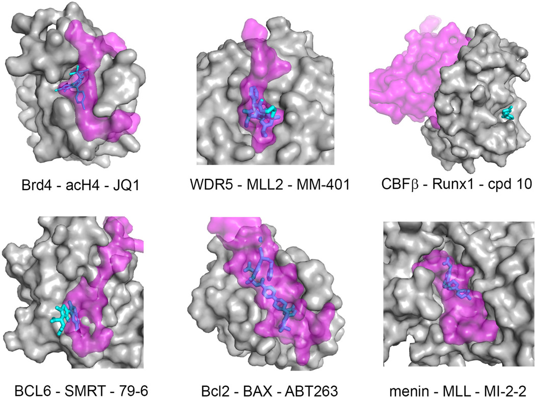
Fig. 3. Comparison of protein-small molecule contacts with protein-protein (or protein-peptide) interaction interfaces.
Target protein is shown in surface representation (gray), protein (or peptide) binding partner is shown in semi-transparent surface (magenta) and inhibitors are shown as sticks (cyan/blue). Blue color corresponds to the region of the ligand molecule that overlaps with binding of the protein (peptide) partner, while cyan color corresponds to the ligand portion that does not overlap with binding of the protein (peptide) partner. PDB codes for PPI complexes are as follows: Brd4-acH4 (3UVW), WDR5-MLL2 (3UVK), BCL6-SMRT (1R2B), Bcl-2-BAX (2×A0); menin-MLL (3U88); CBFβ-Runx1 (1H9D). The PDB structures for protein-inhibitor complexes are the same as shown in Fig. 2.

