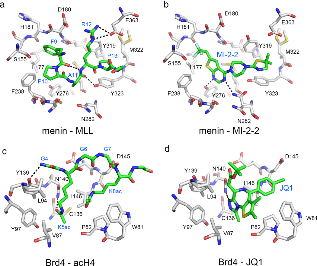
Fig. 5. Comparison of binding modes of natural protein partners and small molecule inhibitors of PPIs.
Details of the interaction of MLL derived peptide (A) and MI-2-2 (B) with menin, demonstrating that MI-2-2 occupies the same region of the binding site and closely mimics key interactions of MLL (in particular residues F9 and P13) with menin (PDB codes: 4GQ6 and 4GQ4, respectively). Comparison of the binding mode of acetylated H4 peptide (D) and JQ1 (D) to Brd4 (PDB codes: 3UVW and 3MXF, respectively). Protein residues, peptides and small molecules are shown in stick representations, with carbon atoms in gray (proteins) or green (peptides and small molecules). Color coding for other heavy atoms remains the same for all complexes: oxygens in red, nitrogens in blue, sulfur in yellow and fluorines in cyan. Dashed lines correspond to hydrogen bonds.

