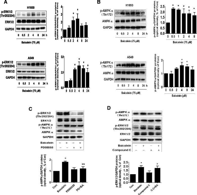
Figure 3.

Baicalein increased the phosphorylation of ERK1/2 and AMPKα in a time-dependent manner. A-B, A549 and H1650 cells were treated with baicalein (75 μM) in the indicated times, and cell lysate was harvested and the expression of the phosphorylated or total protein of ERK1/2, AMPKα were measured by Western blot analysis using corresponding antibodies. GAPDH was used as loading control. C-D, H1650 cells were treated with PD98059 (10 μM) (C) or compound C (C.C, 5 μM) (D) for 2 h before exposure of the cells to baicalein (BA, 75 μM) for an additional 24 h. Afterwards, the expression of p-ERK1/2 and p-AMPKα protein and their total forms was detected by Western blot. The bar graphs represented the densitometry results of p-ERK or AMPKα /GAPDH as mean ± SD of at least three separate experiments. *indicates significant difference from untreated control cells (P < 0.05). **indicates significant difference from baicalein treated alone (P < 0.05).
