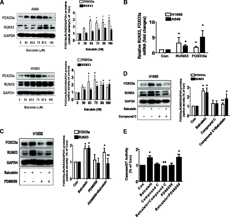
Figure 4.

Baicalein increased protein levels of FOXO3a and RUNX3 through ERK1/2 and AMPK pathways. A, H1650 and A549 cells were exposed to increased concentration of baicalein for 24 h. Afterwards, the expression of FOXO3a and RUNX3 protein were detected by Western blot. B, H1650 and A549 cells were exposed to baicalein for 24 h. Afterwards, the expression of FOXO3a and RUNX3 mRNAs was detected by real-time RT-PCR method as described in the Materials and Methods section. C-D, H1650 cells were treated with PD98059 (10 μM) (C) or compound C (5 μM) (D) for 2 h before exposure of the cells to baicalein (75 μM) for an additional 24 h. Afterwards, the expression of FOXO3a and RUNX3 protein were detected by Western blot using antibodies against FOXO3a and RUNX3. The bar graphs represent the mean ± SD of RUNX3/GAPDH and FOXO3a/GAPDH of three independent experiments. E, H1650 cells were treated with PD98059 (10 μM) or compound C (5 μM) for 2 h before exposure of the cells to baicalein (75 μM) for an additional 24 h. Afterwards, the cells were collected and processed for analysis of apoptosis as determined by caspase 3/7 activity assays. Results represent those obtained in three experiments. *indicates significant difference as compared to the untreated control group (P < 0.05). **indicates significant difference from baicalein treated alone (P < 0.05).
