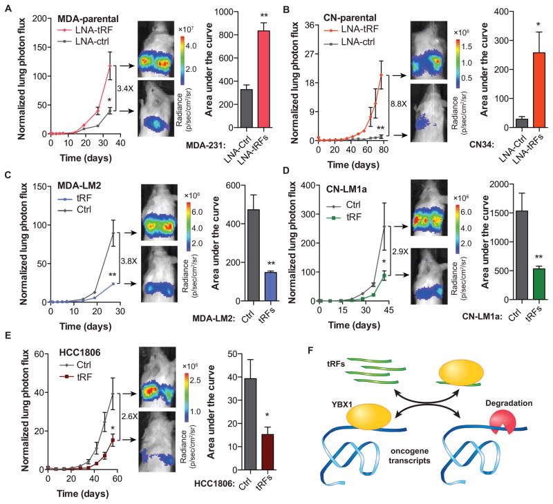
Figure 7. YBX1-binding tRFs play a significant role as suppressors of tumor progression and metastasis.
(A–B) Bioluminescence imaging plot of metastatic lung colonization by MDA-parental and CN-parental cells transfected with synthetic antisense LNAs against all four YBX1-binding tRFs. Representative images along with quantification of the area-under-the-curve for each mouse are also included (n=3–5 in each cohort). (C–D) Bioluminescence imaging plot of lung metastasis by MDA-LM2 and CN-LM1a cells transfected with the four YBX1-binding tRFs. Representative images and area-under-the-curve quantifications are also included (n=4–5 in each cohort). (E) Bioluminescence imaging plot of lung metastasis by HCC1806 cells transfected with the four YBX1-binding tRFs. Representative images and area-under-the-curve quantifications are also included (n=5 in each cohort). (F) Schematic of tRF-mediated modulation of invasion and metastatic lung colonization through in vivo titration of YBX1 and the subsequent destabilization of its oncogenic and pro-metastatic targets. For comparing metastasis colonization assays, two-way ANOVA was used to measure statistical significance. For all other cases, statistical significance is measured using one-tailed Student’s t-test: *, p<0.05, **, p<0.01, and ***, p<0.001. Error bars in all panels indicate s.e.m. unless otherwise specified.

