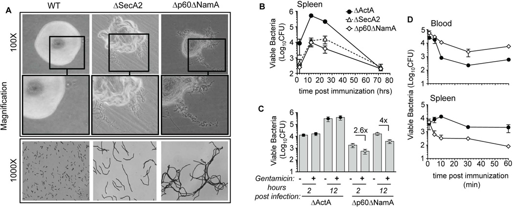
Figure 2. Lm lacking p60 and NamA autolysins exhibit a filamentous phenotype promoting extracellular growth within infected spleens.
(A) Morphology of Lm colonies grown OVN onto BHI Petri dishes, and corresponding gram staining of bacteria. (B–D) WT C57BL/6 (B6) mice were infected with 0.1×LD50 ΔActA (106), ΔSecA2 (0.6×10⁶) and Δp60ΔNamA (0.6×10⁷) Lm; spleen harvested at the indicated times, lysed and plated. In (C), mice received gentamycin i.p. (2 mg) 1 hr after Lm infection. In (D) immunized mice were bled (200µl/mouse), spleens harvested at indicated times, and Lm CFU determined by plating both cell suspensions after lysis. Data plots and histogram bars average mean values+/−SEM (B-D).

