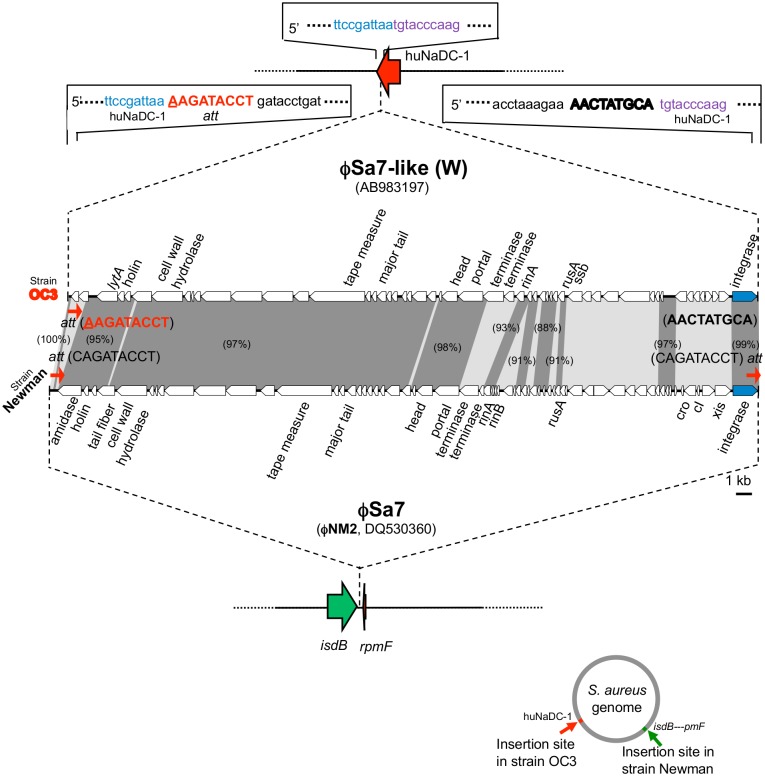
Fig 4. Structure of a phage φSa7-like (W) of the ST239Kras strain OC3.
φSa7-like (W) was compared with φSa7 of the strain Newman for the phage structure, the integration site (att) sequence, and integration site. Homologous regions are shaded in the comparison. The figure at the lower right side indicates each integration site on the S. aureus chromosome. The target huNaDC-1 sequence of φSa7-like (W), shown at the top of the figure (in blue and purple), was also present in the huNaDC-1 gene of other S. aureus strains.

