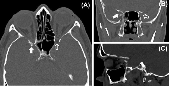
Fig. 4.

Optic canal fracture and optic nerve compression. (A) Axial computerized tomography (CT) scan through the optic nerves showing a fracture in the posterior part of the lateral wall of the right orbit. The bone fragment is causing compression of the right optic nerve within the optic canal (white arrow). A normal wide optic canal can be seen on the left side (clear arrow). (B) Coronal CT scan showing narrowing of the right optic foramen (white arrow) compared with the left side (clear arrow). (C) Sagittal CT scan showing compression of the right optic nerve by the bone fragment. (Courtesy of Dr Manjunatha YC, Sri Devaraj Urs Medical College, Tamaka, Kolar, India.)
