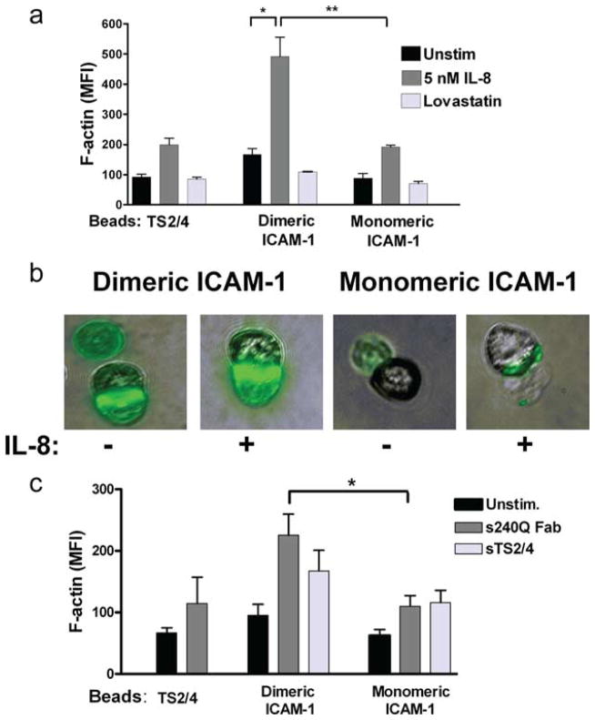
FIGURE 2.
Increase in F-actin expression upon binding of beads coated with ICAM-1. Whole-cell F-actin in response to PMN capture of beads coated with anti-LFA-1 TS2/4, monomeric ICAM-1, or dimeric ICAM-1 as measured by phalloidin-FITC binding and detected by flow cytometry in the presence of (a) soluble IL-8 (5 nM) or lovastatin (1 mM). Data are expressed as MFI from three to five separate experiments. *, p < 0.01 and **, p < 0.001. b, Immunofluorescence images depict, from left to right: phalloidin signal from dimeric ICAM-1 beads; dimeric ICAM-1 coexpressed with IL-8; monomeric ICAM-1; monomeric ICAM-1 with IL-8. c, In the presence of soluble anti-CD18 (240Q Fab, 10 μg/ml) or anti-LFA-1 (TS2/4, 10 μg/ml). *, p < 0.01. Statistical analysis also reported significance in sTS2/4-mediated activation of PMN bound to both dimeric and monomeric ICAM-1 (p < 0.05).

