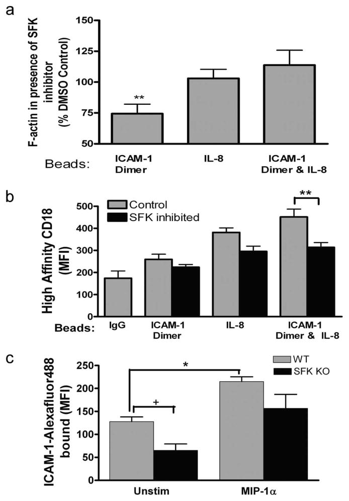
FIGURE 3.
SFK regulation of CD18 affinity and outside-in signaling. Expression of F-actin and high-affinity CD18 were assayed using flow cytometry of PMN/bead complexes. a, Total cellular F-actin formation after treatment with Src kinase inhibitor II (10 μM) during PMN binding to beads coated with dimeric ICAM-1, IL-8, or beads expressing both dimeric ICAM-1 and IL-8. Data are expressed as the percentage of the phalloidin MFI of DMSO-treated control PMN binding to the respective bead type from three to five separate experiments. **, p < 0.05 for difference from 100%. b, Expression of high-affinity CD18 on DMSO control and SFK inhibitor (10 μM)-treated PMN as detected by mAb 327C. PMN were agitated with beads coated with IgG control, dimeric ICAM-1, and dimeric ICAM-1 and IL-8 during three to five separate experiments. **, p < 0.001. c, Soluble dimeric ICAM-1 was conjugated to Alexa Fluor 488 and its binding to mouse wild-type (WT) and lyn−/− hck−/− fgr−/− (SFK KO) PMN in the presence and absence of MIP-1α (100 ng) was quantified by MFI from three separate experiments. *, p < 0.01 and +, p < 0.05. MIP-1α enhanced ICAM-1 binding in SFK KO with a significance level of p = 0.05.

