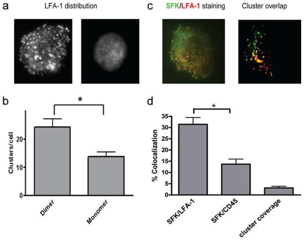
FIGURE 5.
LFA-1 clusters are associated with activated SFK. PMN were allowed to settle on ICAM-1-bearing glass coverslips and then fixed, stained, and imaged for LFA-1 and phospho-SFK distribution. a, Representative images of LFA-1 distribution on settled neutrophils were enhanced using gamma adjustment to demonstrate the distribution of high-density clusters. b, The number of clusters per neutrophil was quantified by directly counting the number of distinct regions with fluorescent pixel intensity 2.5 SDs higher than the average. *, p < 0.01. c, PMN were plated on ICAM-1 and then fixed and stained with Alexa Fluor 488-conjugated anti-LFA-1 Ab followed by permeabilization and staining with anti-phospho-SFK (Tyr416) as in a. PMN were imaged by total internal reflection fluorescence microscopy, and colocalization of LFA-1 and Tyr416 or of CD45 and Tyr416 was quantified by rendering clusters of LFA-1, CD45, and Tyr416 into pixilated bimodal images as shown. d, Colocalization was defined as the percentage of Tyr416 pixels that coincided with LFA-1 or CD45 pixels, with “+” denoting significance between monomeric and dimeric ICAM-1 and LFA-1 and CD45 at p < 0.05.

