
Figure 1.
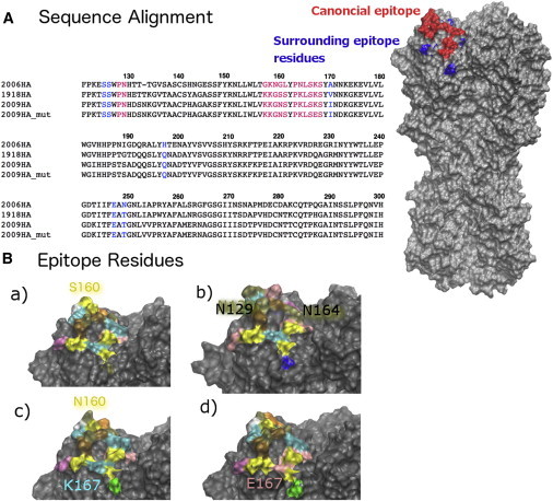
Sequence alignment and the epitopes of the four HA glycoprotein. (A) Sequence alignment and structural view. The four HA sequences are aligned and numbered using the 18HA numbering convention. On the right, the Sa epitope is colored red and the surrounding residues that form contact with Ig-2D1 (12) are colored blue (top left) in 18HA monomer 1. (B) Structural conservation of the HA epitope region and key mutations that affect antibody recognition. The four HAs are shown: (a) 18HA, (b) 06HA, (c) 09HA, and (d) 09HA_mut. The epitope residues on monomer 1 are colored by residue names. Several key residues are also labeled to their corresponding residue colors. S160 (18HA) is mutated in N160 in 09HA. K 167 (09HA) is mutated to E167 in 09HA_mut. N129 and N164 in 06HA are potential glycosylation sites. To see this figure in color, go online.