
Figure 3.
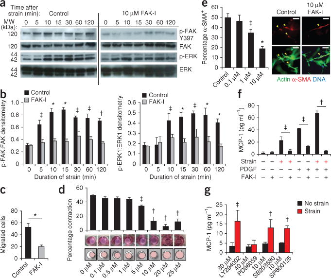
FAK-mediated mechanoresponsive pathways in human fibroblasts. (a,b) Representative immunoblots and quantification of static strain-induced FAK and ERK activation in untreated human fibroblasts (Control) and those treated with the FAK inhibitor (FAK-I) PF573228. n = 3. (c) Fibroblast motility in a scratch migration assay. n = 6. (d,e) Fibroblast contraction (d) and α-SMA+ expression (arrowheads) (e) in three dimensional collagen lattices. Concentrations of PF573228 are indicated along the x-axes of the bar graphs. n = 3. Scale bars, 50 μm. (f) Synergistic (strain plus 10 ng ml−1 platelet-derived growth factor) induction of MCP-1 secretion. n = 4. (g) Strain-induced MCP-1 secretion with small-molecule inhibition of FAK (PF573228), Akt (LY294002), ERK (PD98059), p38 (SB203580) or JNK (SP600125). n = 4. Values are means ± s.e.m. *P < 0.001, †P < 0.01, ‡P < 0.05.