
Fig. 1. Generation of the Nrmt1−/− knockout mouse line.
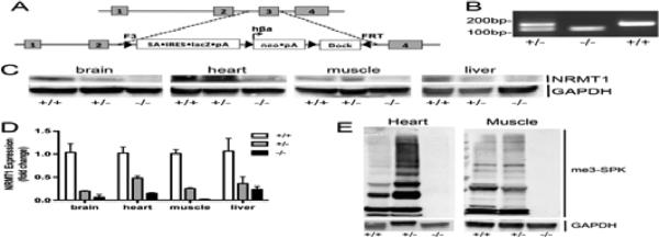
(a) Diagram of the reporter construct used to replace the third exon of NRMT1 and generate the Nrmt1−/− mice. (b) PCR genotyping of Nrmt1+/− heterozygotes (+/−), Nrmt1−/− homozygotes (−/−), and wild type C56BL/6J (+/+) mice. (c) Western blot illustrating loss of NRMT1 protein expression in Nrmt1−/− brain, heart, muscle, and liver tissue as compared to wild type and heterozygotes. GAPDH is used as a loading control. (d) qRT-PCR analysis of NRMT1 mRNA levels in wild type, heterozygous, and homozygous Nrmt1−/− mouse tissues. Levels given as fold change from wild type. (e) Western blot illustrating that along with NRMT1 protein levels, there is a loss of N-terminally methylated proteins (me3-SPK) in the Nrmt1−/− heart and muscle tissue as compared to wild type and heterozygotes. GAPDH was used as a loading control.