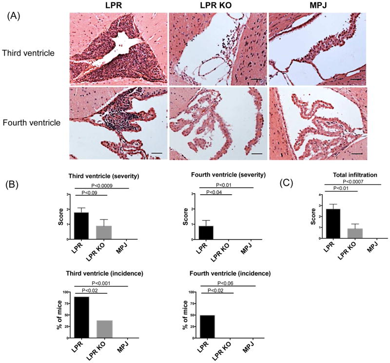
Figure 1.
Cellular infiltration in the brains of MRL/lpr Fn14WT (LPR), MRL/lpr Fn14KO (LPR KO), and MRL/MPJ (MPJ) mice. H&E staining (A) was used to assess the cellular infiltration in the third and fourth ventricles of LPR, LPR KO, and MPJ mice at 20 weeks of age. Quantification of the severity and incidence of infiltration is shown in (B). The total infiltration score of each mouse was calculated in (C). The number of mice in the LPR, LPR KO, and MPJ groups was 10, 8, and 5, respectively. The scale bars in these and all subsequent figures indicate 50 μm.

