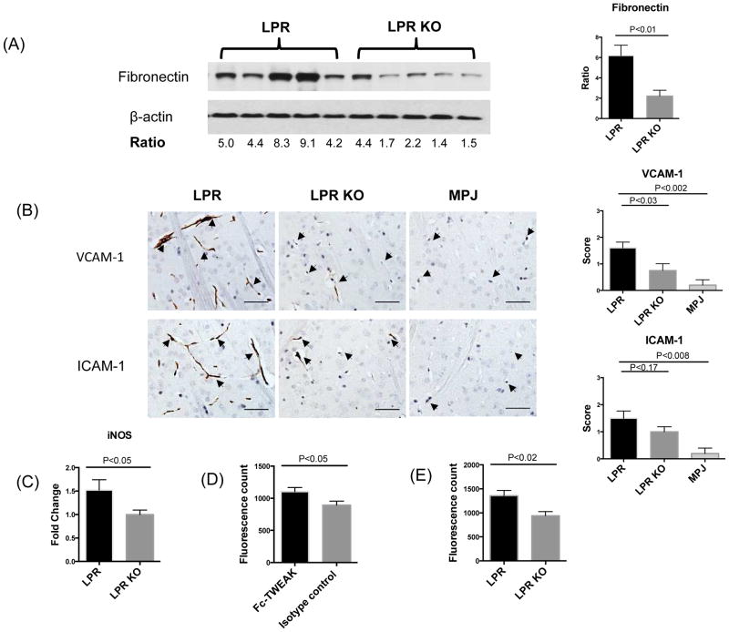
Figure 3.
BBB integrity in LPR and LPR KO mice. Western blot for fibronectin (A), left panel, was performed on brain lysates from randomly selected LPR (n=5) and LPR KO (n=5) at 20 weeks of age. Each band represents one individual mouse. Quantification of fibronectin expression by Image J is shown in (A), right panel. VCAM-1 and ICAM-1 staining is shown in (B), left panel. Arrows indicate endothelial cells. Staining intensity was graded semi-quantatively from 0 (absent staining) to 3 (maximal staining) (B), right panel. The number of mice for VCAM-1/ICAM-1 staining in the LPR, LPR KO, and MPJ groups was 10, 8, and 5, respectively. (C) iNOS was detected by qRT-PCR in cortical mRNA samples from LPR (n=8) and LPR KO (n=8) mice. The fluorescence intensity in the supernatants of brain lysates from MRL/lpr mice treated with Fc-TWEAK (n=5) or control IgG (n=5) is shown in (D). The fluorescence intensity in the supernatants of brain lysates from MRL/lpr Fn14WT (n=5) and Fn14KO mice (n=5) treated with Fc-TWEAK mice is shown in (E). Western blot for fibronectin was repeated three times, and VCAM-1/ICAM-1 staining and qPCR for iNOS were repeated twice, with similar results. The Fc-TWEAK injection experiments were done once.

