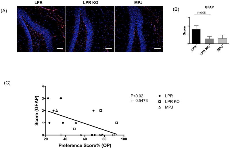
Figure 7.
Hippocampal gliosis correlates with spatial memory in LPR and LPR KO mice. GFAP staining of LPR (n=9), LPR KO (n=8), and MPJ (n=5) at 20 weeks of age is shown in (A). Representative images of each group are shown in the figure. A semi- quantitative gliosis score based on GFAP staining (B) was as follows: 0=no gliosis, 1=low level of gliosis, 2=moderate level of gliosis, and 3=high level of gliosis. GFAP staining was repeated twice, with similar results. Linear regression correlation of the scores of hippocampal gliosis with the preference scores in the object placement test is shown in (C).

