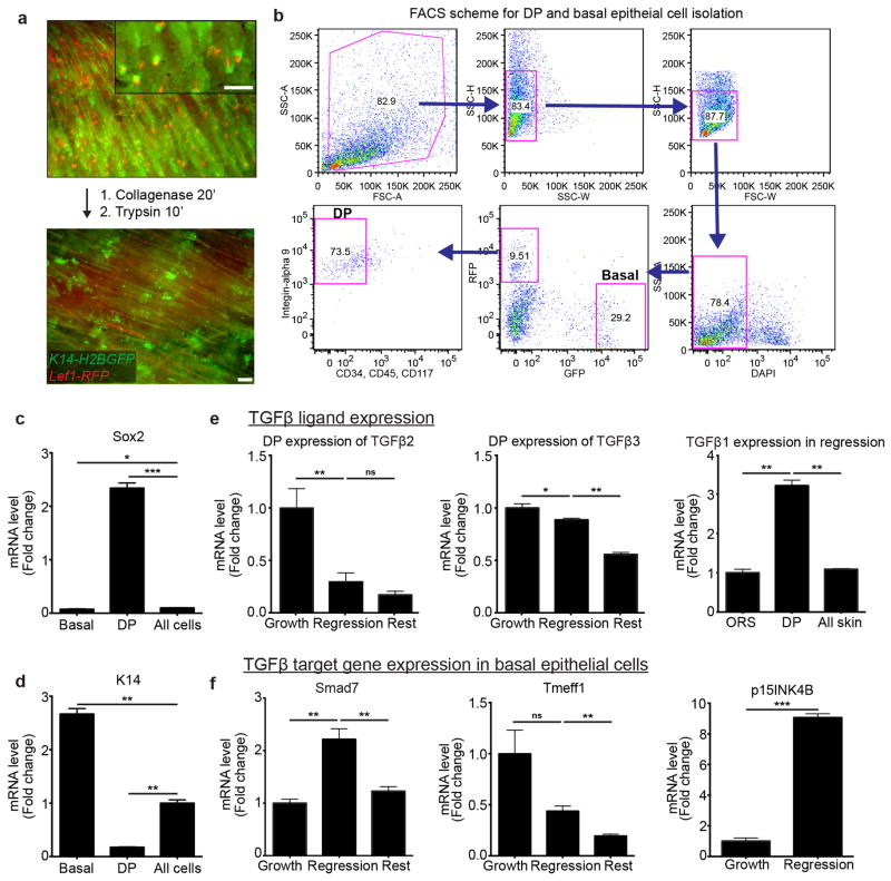
Extended Data Fig. 7. Characterization of TGFβ pathway in mesenchymal DP and basal epithelial cell populations during regression.
a) Schematic of skin digestion and cell isolation with representative images before and after tissue digestion, in K14-H2BGFP;Lef1-RFP mice. b) Representative FACS scheme for isolating mesenchymal DP (RFP+, CD34−, CD45−, CD117−, Integrin-alpha 9+) and enriched hair follicle basal epithelium (RFP−, GFPHigh) cells. c) Validation of mesenchymal DP sorted population enrichment by Sox2 expression. d) Validation of basal epithelial sorted population enrichment by keratin-14 (K14) expression. e) TGFβ ligand 2 and 3 expression in the mesenchymal DP throughout the hair cycle. TGFβ-1 expression in basal epithelium, mesenchymal DP, and all sorted cells during regression. f) Differential expression of TGFβ target genes: Smad7, Tmeff1, p15INK4B and in the hair follicle basal epithelium throughout the hair cycle (p<0.005 mean ± SD; n = 3 technical replicates). Scale bars, 100μm.

