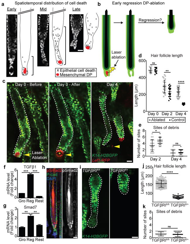
Figure 3. Mesenchymal DP crosstalk and TGFβ signaling are required for cell death in the basal epithelium.
a) Graphical representation and quantification of spatial distribution of cell death in the basal layer at three stages of regression using time-lapse recordings (n = 9 follicles, in 5 mice). b) Scheme of laser ablation experiment. c) Sequential revisits of hair follicles after DP ablation during regression. Yellow arrowhead indicates laser ablation site. Asterisk indicates auto-fluorescence from the two-photon laser. d) Dot plot quantification of the hair follicle length at Day 0, 2 and 4 after DP ablation, and e) of the number of apoptotic fragmentation sites at Day 2 and 4 after DP ablation (n = 36 follicles, in 6 mice; mean ± SD). f) mRNA levels of TGFβ1 ligand expression in the mesenchymal DP and g) Smad7 expression in hair follicle basal layer throughout the hair cycle (p<0.005; mean ± SD; n = 3 technical replicates). h) Localized TGFβ activation by immunofluorescent staining of pSmad2 at the onset of hair follicle regression. DAPI (blue), pSmad2 (red), and P-cadherin (green). i) Hair follicles after Cre-induced recombination in TGFβR1fl/fl and TGFβR1fl/+ mice at the end of regression. j) Dot plot quantification of the hair follicle length, and k) of the number of apoptotic fragmentation sites at the end of regression (n = 31 follicles, in 4 mice; mean ± SD). Scale bars, 25 μm.

