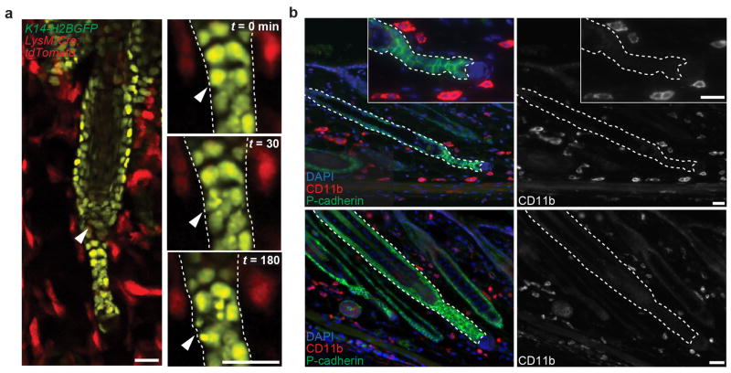
Extended Data Fig. 4. Professional phagocytes are not present in regressing hair follicles.
a) Professional phagocytes do not enter regressing hair follicles. Single optical sections showing absence of myeloid populations inside the hair follicle 2.5 h after epithelial cell death (arrowhead), in LysM-Cre; tdTomato;K14-H2BGFP mice. b) Immunofluorescent staining of myeloid populations in skin during hair follicle regression. DAPI (blue), CD11b (red), and P-cadherin (green). Observations shown represent n = 4 mice. Scale bar, 25 μm

