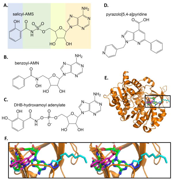
Figure 5. Inhibitors of salicylate and dihydroxybenzoate adenylation enzymes.
A. Salicyl-AMS, a rationally designed reaction intermediate analogue. [36]. B. Benzoyl-AMN [43]. C. DHB-hydroxamoyl adenylate [44]. D. Inhibitor from high throughput screen. [46]. E. Three structures of BasE are overlayed: BasE with DHB-AMS bound is the orange cartoon with green stick inhibitor (PDB ID: 3O82), with a triazole derivative of DHB-AMS shown in cyan sticks (PDB ID: 3O83), and with inhibitor from part D shown in magenta sticks (PDB ID: 3O84). F. The area in the box in part E is shown in stereo (all stereo images are wall-eye or divergent stereo). Note the unexpected binding mode of the magenta HTS inhibitor relative the green and cyan substrate analogue inhibitors. Structure figures made in PyMOL [121].

