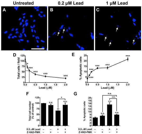
Fig. 2. Lead significantly decreases the total cell number and increases apoptosis in SGZ-aNPCs.
A-E, SGZ-aNPCs were treated with 0, 0.1, 0.2, 0.5, 1, or 2 μM lead for 48 h. Representative Hoechst nuclei staining from (A) untreated, (B) 0.2 μM, and (C) 1 μM lead-treated SGZ-aNPCs. Quantification of (D) the total cell number and (E) the percent apoptotic cells. F-G, SGZ-aNPCs were pretreated with 5 μM Z-VAD-FMK, a pan-caspase inhibitor, for 2 h followed by 0.5 μM lead for 48 hand (F) the total cell number and (G) the percent apoptotic cells were quantified. Arrows: nuclear condensat ion and/or fragmentation. Hoechst: nuclei staining. Scale bar: 50 μm. n = 2-3 independent experiments for a total of 4-8 coverslips per data point. Data represent mean ± SEM., n.s. not significant, * p < 0.05; ** p < 0.01; *** p < 0.001.

