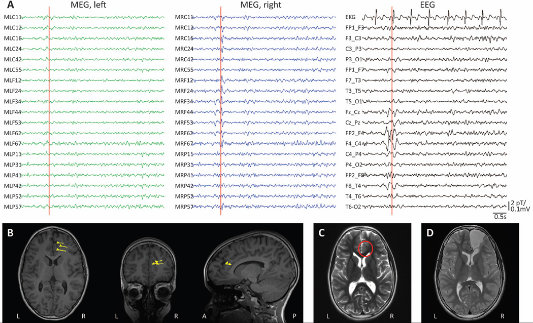
Figure 1. Example of MSI dipole modelling with simultaneous MEG/EEG recordings.
A) Recordings from selected MEG channels and simultaneous EEG in a 12-year-old female with drug-resistant focal epilepsy. A representative interictal spike is seen in both MEG and EEG recordings localizing to the right frontal/central region. B) Localization of single dipole sources corresponding to the spike in A, and similar spikes during the recording, shown as triangles with vector tails superimposed on T1-weighted anatomical MRI. C) Pre-operative T2-weighted axial MRI showing a subtle abnormal blurring of gray-white matter differentiation in the right anterior cingulate region (circle), proximal to the location of MEG dipoles. D) Post-operative T2-weighted axial MRI demonstrating the resection cavity. Neuropathological examination revealed FCD type IIA, and the patient remains seizure-free two years after surgery. EEG: electroencephalography; FCD: focal cortical dysplasia; MEG: magnetoencephalography; MRI: magnetic resonance imaging; MSI: magnetic source imaging.

