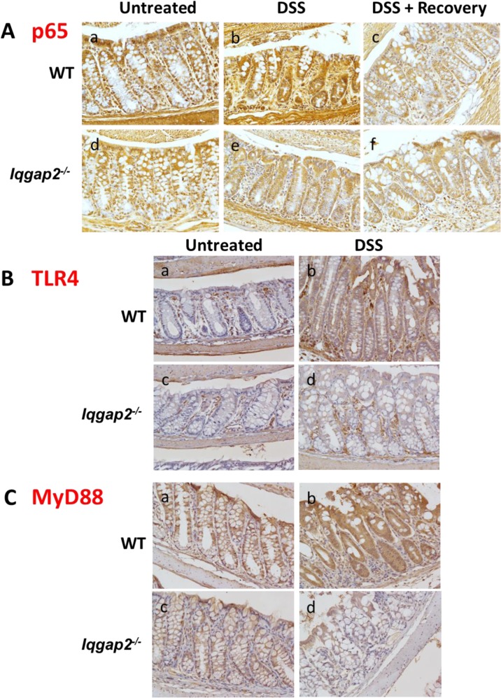
Fig 3. Suppression of NF-κB signaling in Iqgap2 -/- colons.
A. IHC shows decreased baseline levels of p65 subunit of NF-κB in Iqgap2 -/- colon compared to WT (panels a, d). While DSS treatment resulted in elevated levels of p65 in WT colon, it failed to elicit the same response in Iqgap2 -/- colon (panel b, e). Termination of DSS treatment results in a restoration of the baseline p65 levels within 7 days in both genotypes (panel c, f). B. IHC of TLR4 in WT and Iqgap2 -/- colons before and after DSS treatment. C. IHC of MyD88 in the same samples. Note low levels of both TLR4 and MyD88 in Iqgap2 -/- colons after DSS exposure (panels d). A representative image of N = 5 per genotype is shown for each IHC. Magnification is 200 X.

