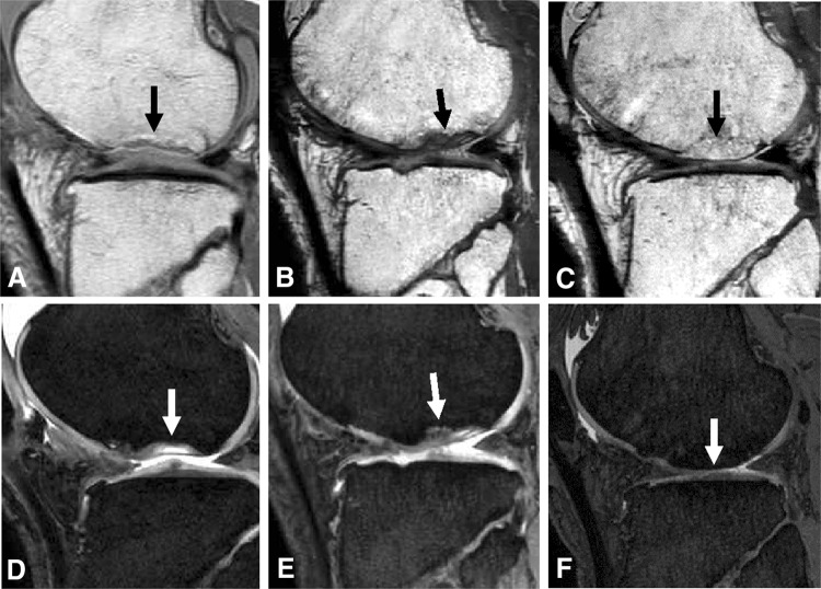
Fig. 4A–F.
Sequential MRI features of Patient 2 who had an osteochondral defect, indicated by the arrow, are shown (A) for bone before treatment, (B) at 6 months, and (C) at 6 years, and for (D) cartilage before treatment, (E) at 6 months, and (F) at 6 years. The sequential MR images showed that the osteochondral defect was filled with cartilaginous tissue at 6 months. The bone defect was filled with bony tissue, and the cartilage defect was incompletely filled with cartilaginous tissue at 6 years.

