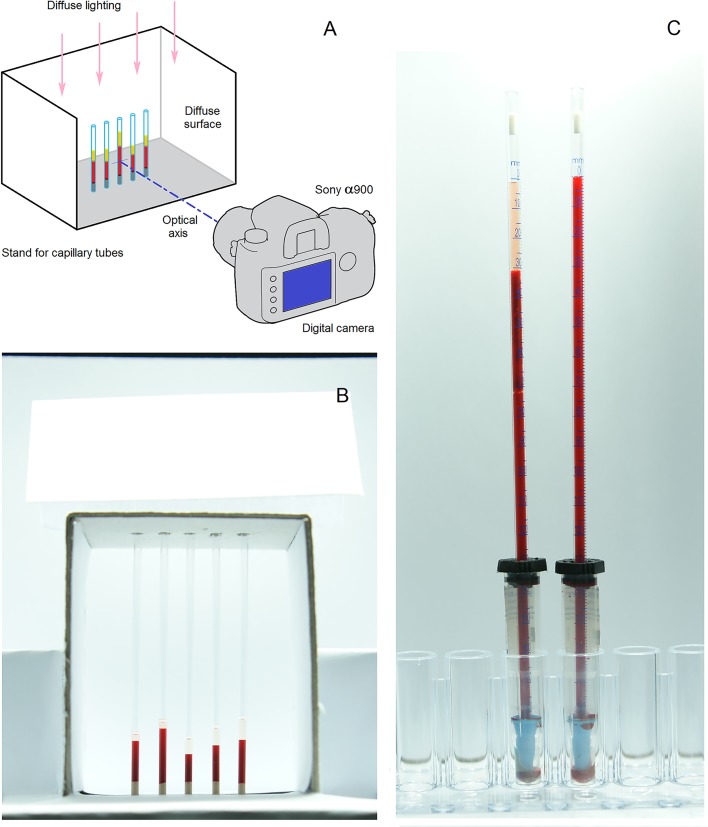
Fig 2. Schematic of the experimental setup that was used to obtain optical observations of blood sedimentation.
(A) Sony α900 digital camera with Carl Zeiss Vario-Sonnar lens. (B) Photograph of the micro-hematocrit capillary tubes in 5-place rack. Each capillary tube has an inner diameter of 1.2 mm and a length of 75 mm. (C) Photograph of the Sedi-Rate ESR System inside the light-tight box. Each plastic tube has a sedimentation scale of 200 mm and a bore of 2.55 mm.

