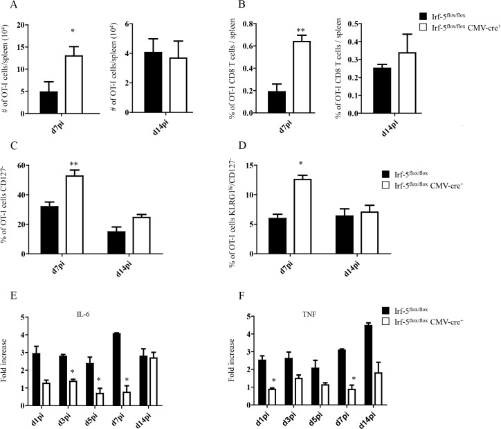
Fig 1. IRF-5-mediated inflammation limits CD8+ T cell expansion during acute L. donovani infection.
(A-D) 2x10⁴ OT-I CD8+ T cells were adoptively transferred into recipient mice a day prior to infection with ovalbumin-transgenic (PINK) L. donovani amastigotes. (A) Graph represents the average number of OT-I CD8+ T cells found in the spleen from Irf-5 flox/flox CMV-Cre + and Cre - mice at d7 and d14 p.i.. (B) Percentage of OT-I CD8+ T cells at d7 and d14 post infection. (C) Percentage of gated OT-I CD8+ that were negative for CD127. (D) Percentage of OT-I CD8+ T cells that did not express CD127 and are positive for KLRG1. Real-time PCR analysis of IL-6 (E) and TNF (F) expression in CD11c+ cells from Irf-5 flox/flox CMV-Cre + and Cre - at various time points after infection. All data represent mean ± SEM of one of 3 independent experiments, n = 5. * denotes p<0.05, ** denotes p<0.01 and *** denotes p<0.001.

