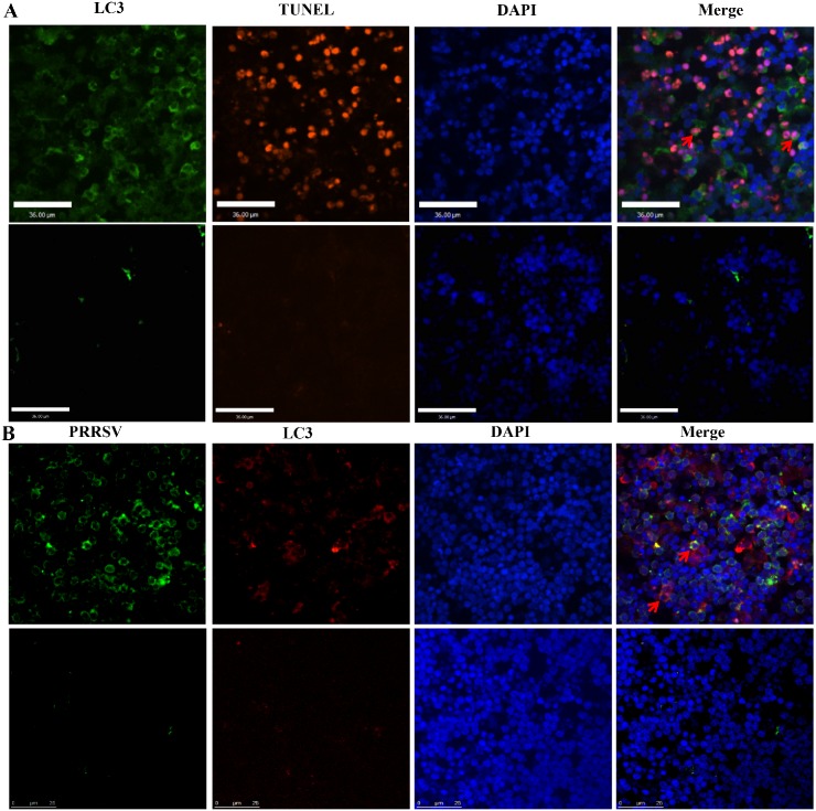
Fig 3. Colocalization of PRRSV-infected cells, autophagic cells, and nuclei.
Autophagic cells were stained with anti-LC3 antibodies and anti-rabbit secondary antibodies conjugated to FITC, and apoptotic cells were detected according to the manufacturer’s instructions for the In Situ Cell Death Detection Kit, only a few apoptotic cells underwent autophagy (arrows) (A); Autophagic cells were stained with anti-LC3 antibodies and tetramethylrhodamine- isothiocyanate (TRITC)-conjugated goat anti-mouse antibodies, and HuN4-infected cells were strained with fluorescein isothiocyanate (FITC)-conjugated monoclonal antibodies (mAbs) against PRRSV N protein, and some HuN4-infected cells underwent autophagy (arrows) (B). Nuclei were stained with 4-6-diamidino-2-phenylindole (DAPI).

