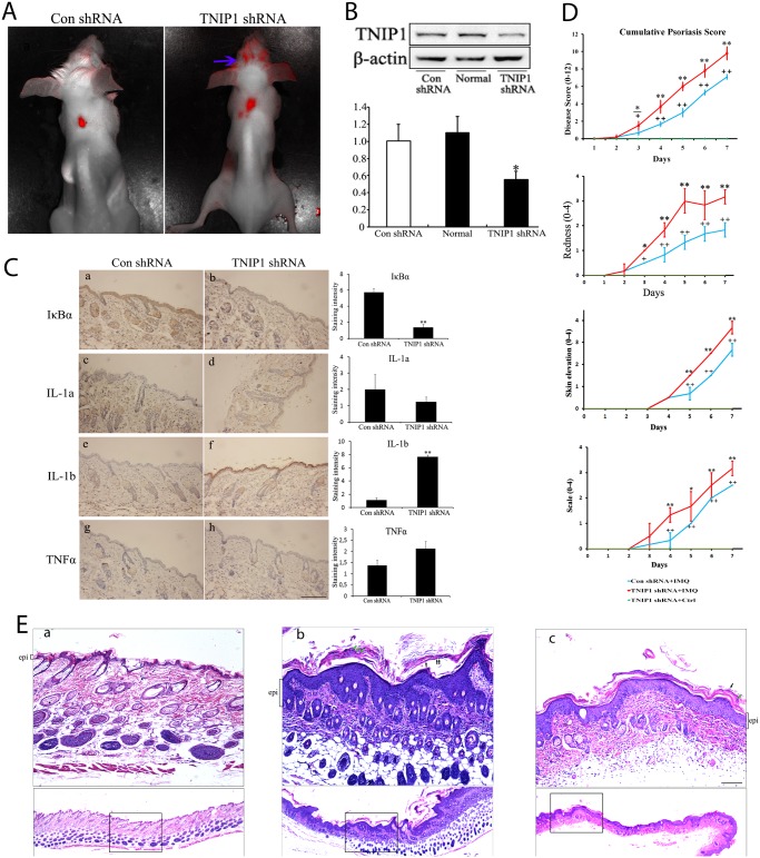
Fig 5. Downregulation of TNIP1 exaggerates IMQ-induced psoriasis-like dermatitis in vivo.
(A) Red fluorescence observed in mice seven days after intradermal injections of lentiviral particles encoding TNIP1 shRNA or control shRNA, respectively. Blue arrows indicate the unspecific fluorescence of mice hair. The back of the mice had been shaved. (B) Western blotting was performed to verify that the TNIP1 expression level was decreased around the injection site. (C) Skin sections obtained seven days after injection from control shRNA mice (n = 4) or from TNIP1 shRNA mice (n = 4) were stained with rabbit anti-IκBα antibody, rabbit anti-TNFα antibody, rabbit anti-IL-1a antibody and rabbit anti-IL-1b antibody, respectively. Bar = 200 μm. Staining intensity was scored in a semiquantitative manner by two independent observers. Data are presented as mean staining intensity grade ± SEM. (D) Clinical scores of IMQ-induced psoriasis-like dermatitis in control shRNA or TNIP1 shRNA-injected mice at the indicated days with or without IMQ treatment. (E) H&E-stained sections of back skin from the mice at the indicated areas treated with or without IMQ. The epithelial layer (epi) is indicated by brackets. Green arrows indicate neutrophil infiltration and black arrows indicate parakeratosis. The epidermal layer thickness was more prominent, but unevenly distributed in TNIP1 shRNA-injected mice (panel b) compared with control shRNA-injected mice (panel c). No obvious epidermal hyperplasia was observed in TNIP1 shRNA-injected mice treated with topical emollient (the control, panel a). Bar = 100 μm. Data are presented as mean ± SD of three independent experiments (* P<0.05 or **P<0.01 compared with controls).

