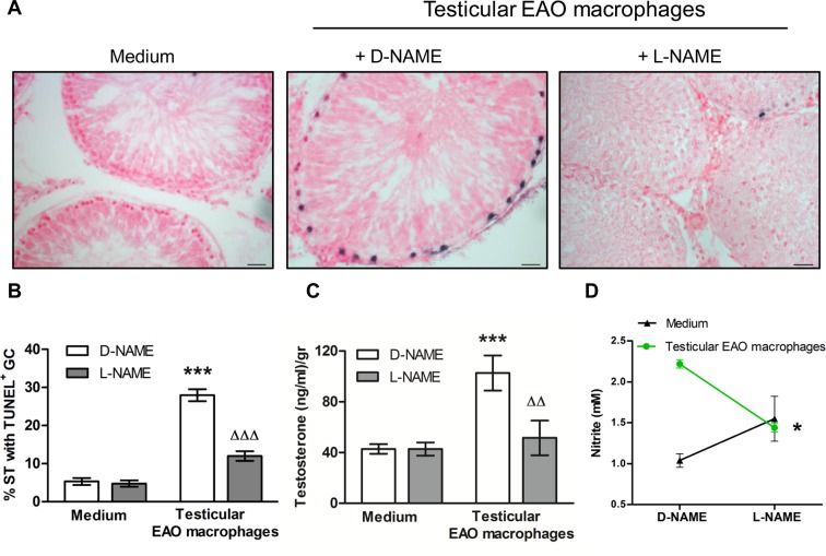
Fig 3. Effect of NO released by testicular macrophages on germ cell (GC) apoptosis (TUNEL technique) and testosterone secretion (RIA).
A) Representative microphotographs of sections of testicular fragments (TF) from normal rats incubated with medium alone or testicular macrophages (TM) isolated from EAO rats in the presence of D-NAME (2mM) or L-NAME (2mM) for 18h; apoptotic GC are blue stained. Scale bar indicates 20μm B) % of seminiferous tubules (ST) with apoptotic GC, the % of ST with apoptotic cells in the TF immediately removed from the testis was 3.140±0.890. Data represent mean±SEM of (n = 30–40) non consecutive sections of TF obtained from four animals in four independent experiments. 300 ST were counted in each condition/rat. ***p<0.001 vs medium D-NAME; ΔΔΔp<0.001 vs D-NAME TM. Two-way ANOVA followed by Bonferroni Multiple Comparison Test. C) Testosterone content in the media. Data represent the mean±SEM (n = 5–7 wells/group from two experiments); ***p<0.001 vs medium D-NAME, ΔΔ p<0.01 vs D-NAME TM. Two-way ANOVA followed by Bonferroni Multiple Comparison Test. D) Nitrite production measured in the medium (fluorometric kit). Data represent mean±SEM (n = 5–7 wells/group from two experiments). *p<0.05 vs D-NAME TM. Student t Test.

