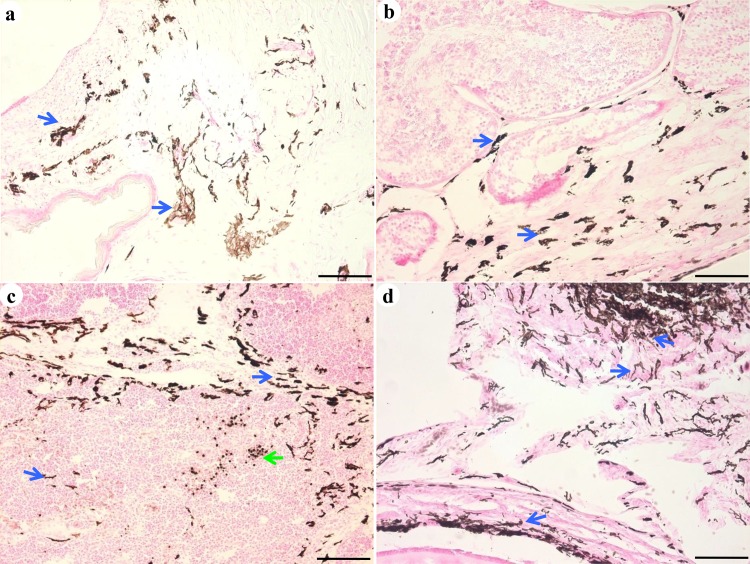
Fig 3. Melanocyte morphology.
Numerous melanocytes (indicated by a blue arrow), with the appearance of dendritic cells and with round nuclei and abundant melanin, were observed in the skin (a), testis (b), thymus (c), and ovary (d). Macrophages in the thymus were stained by 3, 4-dihydroxy-l-phenylalanine (DOPA; c, green arrow). Scale bar = 100 μm.

