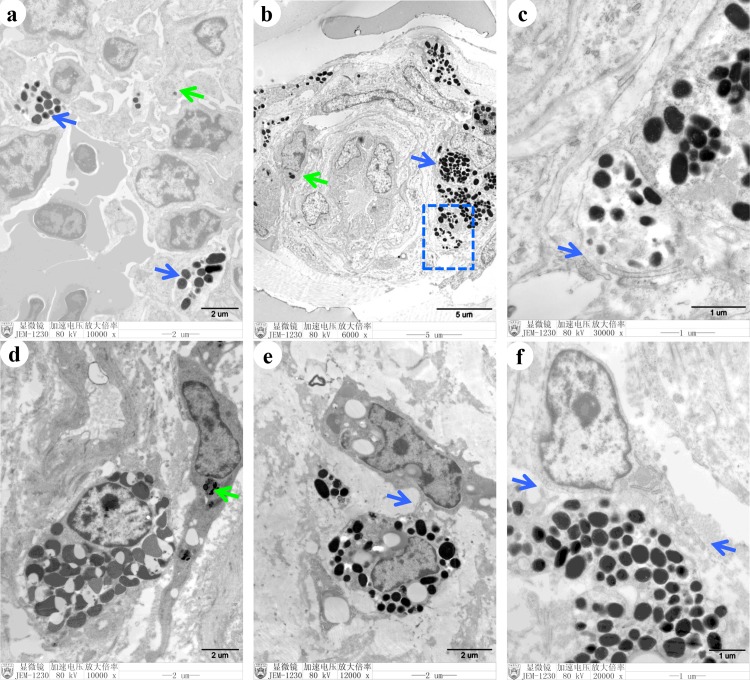
Fig 5. Tissue-specific ultrastructural characteristics of melanocytes and neighboring cells.
Transmission electron microscopy was used to observe the melanocytes. Melanosomes were observed in the melanocytes (blue arrow) and the immune cells (green arrow) of the thymus (a). Melanosomes were present in other cells (green arrow) in the skin (b). Melanosomes were secreted by exocytosis from melanocytes in the skin (c). Melanosomes were observed in the interstitial cells (green arrow) in the ovaries (d). Melanocytes and neighboring cells were connected by dendrites in the ovaries (e). Long dendrites were detected in melanocytes and neighboring cells in the ovaries (f).

