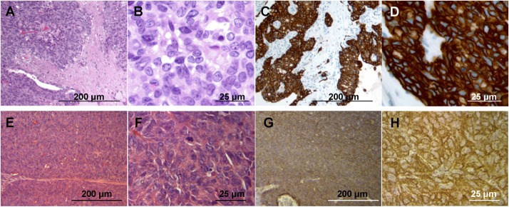
Fig 1. Tumor histology and immunohistochemistry.

(A) H & E-stained section of the original patient tumor. (B) High magnification image of (A). (C) Immunostained section of the original patient tumor using anti-HER-2 antibody. (D) High magnification image of (C). (E) H & E-stained section of the mouse-grown tumor. (B) High magnification image of (F). (G) Immunostained section of the mouse-grown tumor using an anti-HER-2 antibody. (H) High magnification image of (G). Scale bars: 200 μm (A, C, E and G) and 25 μm (B, D, F and H).
