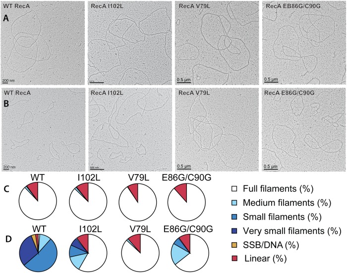
Fig 4. Electron microscopy of RecA variant protein filaments on cssDNA, with and without treatment by RecX protein.
Electron micrographs show filament formation of wild type RecA and RecA variant proteins on M13mp18 ssDNA (A) without RecX protein and (B) with 100 nM RecX protein. RecA filaments were placed into 5 different categories based upon the size and completeness of filaments. (C) The composition of filaments in the various categories without RecX protein and (D) with RecX protein.

