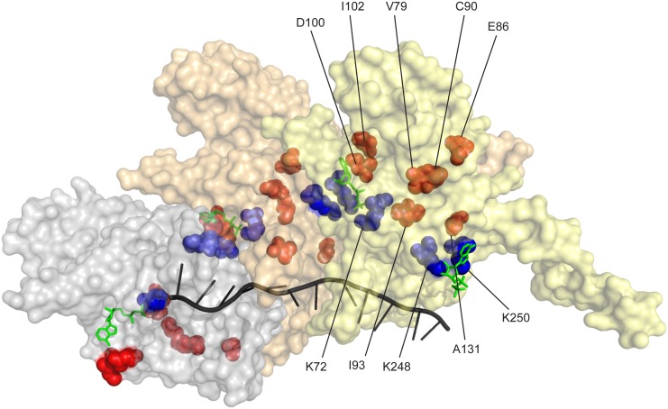
Fig 8. RecA protein amino acid residues affected in RecA variants with increased recombination potential.
Three RecA subunits in a RecA-ssDNA nucleoprotein filament (from coordinates provided by Pavletich and coworkers [165]) are shown, with a surface contour rendering in which each subunit is transparent but differently colored. The path of the ssDNA within the filament is shown by the black helical line. ADP residues are shown in green. The ATPase active site is at the subunit-subunit interface. Three residues at the ATPase active site (K72 on one face and K248 and K250 on the opposing face) are shown in blue. Prominent residues in which amino acid changes bring about enhanced recombination potential are shown in red/orange.

