
Figure 4.
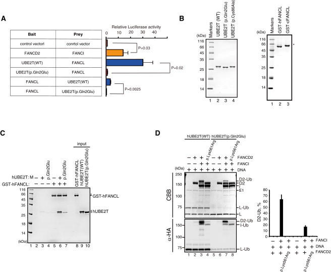
Functional Interaction between FANCL and UBE2T
(A) Mammalian two-hybrid assay. The assays were carried out as described.19 In brief, UBE2T and FANCL in the bait vector (pM) or prey vector (pVP16) were co-transfected into 293T cells with the reporter luciferase vector as well as an internal control (pRL Renilla Luciferase vector). Luminescence signals were quantified using a Dual-Glo Luciferase Reporter Assay System (Promega). The signal was first normalized to transfection efficiency using Renilla luciferase levels and further divided by the value obtained by the empty bait and prey vector. The mean and SD of more than three independent experiments are shown. Statistical analysis was done using Student’s t test, and p values are indicated.
(B) Purified recombinant proteins detected by Coomassie brilliant blue (CBB) gel staining. Human UBE2T proteins with or without the p.Gln2Glu substitution were produced in E. coli and purified to homogeneity as previously described.11 WT, wild-type.
(C) In vitro pull-down assay between GST-human (h)FANCL and hUBE2T protein with or without the p.Gln2Glu substitution. Purified human UBE2T (6 μg) and GST-chicken FANCL or human FANCL (9 μg) were incubated at 37°C for 1 hr in 200 μl of reaction buffer containing 20 mM Tris-HCl (pH 7.5), 10% glycerol, 100 mM NaCl, 1 mM ZnOAc, 0.01% NP-40, and 5 mM 2-mercaptoethanol. Glutathione sepharose 4B beads (3 μl; GE Healthcare) were added to the reaction mixtures, and reaction mixtures were gently mixed at 25°C for 1 hr. The beads were then washed twice with 1 ml of reaction buffer. The proteins bound to the beads were separated by 15% SDS-PAGE and were visualized by Coomassie brilliant blue staining. Asterisk indicates read-through products.
(D) In vitro FANCD2 monoubiquitination assay in the presence of DNA. The assay was repeated three times, and the mean and SD of the percent of monoubiquitinated FANCD2 (D2-Ub) are shown in the graph. Execution of the in vitro FANCD2 ubiquitination reaction was as described.11 As a control, FANCD2 protein carrying a p.Lys561Arg substitution blocking monoubiquitination was included. CBB, Coomassie brilliant blue. Immunoblotting with anti-HA (α-HA) was used to detect HA-tagged ubiquitin.