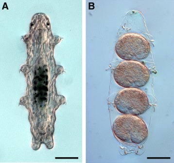
Figure 1.

Habitus of an adult specimen and deposited eggs with embryos of the eutardigrade Hypsibius dujardini. Differential interference contrast (DIC) light micrographs. Anterior is up. (A) A live, adult specimen in dorsal view. The gut full of ingested algae appears as a dark green region in the midbody. (B) A shed cuticle containing four eggs with embryos. Notice the curled shape of each embryo. Scale bars: (A), (B), 50 μm.
