
Figure 8.
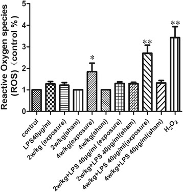
ROS production in primary SGN exposed to RF-EMR and LPS. After exposure to RF-EMR and LPS, SGN were incubated with DCFH-DA at 37°C for 30 min. Cellular fluorescence intensity was expressed as the multiple of the level in control groups. **P < 0.01 compared to the control group, *P < 0.05 compared to the control group. Values are means ± SE, n = 12. LPS, lipopolysaccharide.