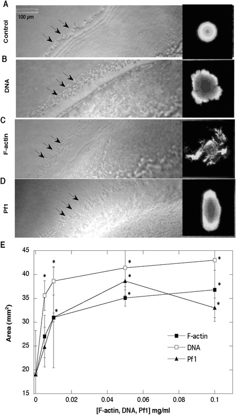
Fig. 5.

Morphology of Pseudomonas aeruginosa Xen5 biofilm growth on agar with addition of DNA, F-actin and Pf1 bacteriophage. In the presence of different biopolymers (0.1 mg/ml), after 16 h of bacterial growth, the swarming edges show some similar features (they are more diffuse and less defined; panel a-d). Developing area of spreading PA biofilm due to swarming activity was assessed using Image Gauge software (panel e). *Significantly different compared to control
