Figure 11.

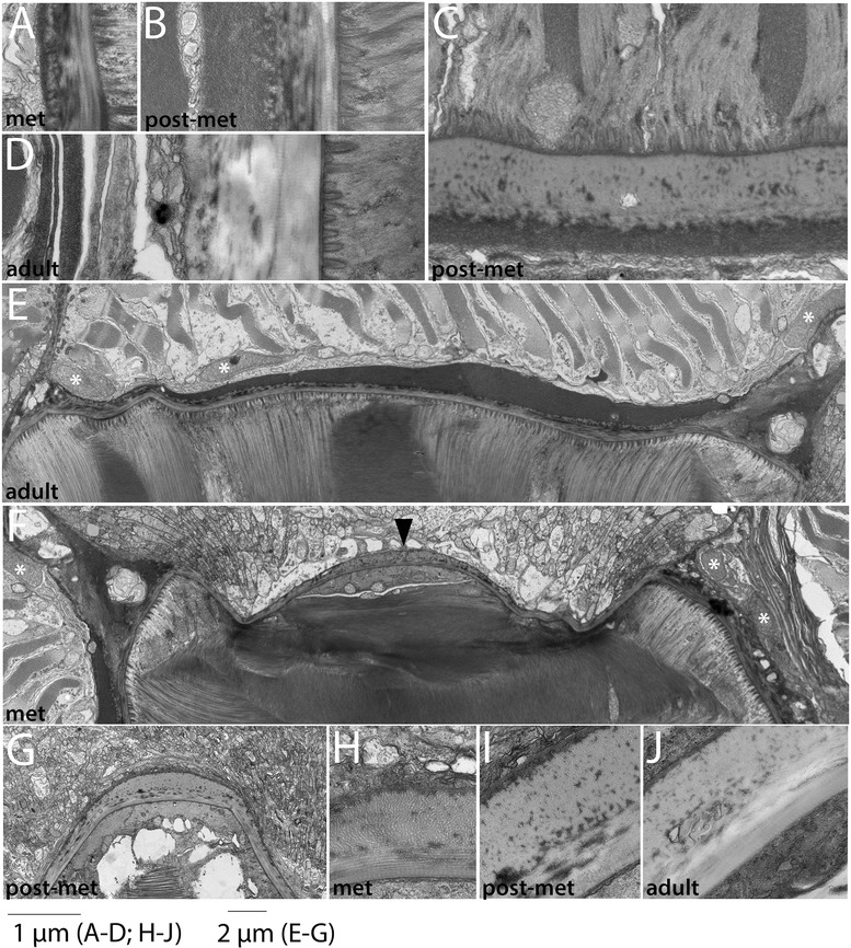
Development of the notochordal sheath (2): metamorphic and adult stages. Striated collagen layers are added to the notochordal sheath through metamorphic and into adult stages. (A) shows the same panel as in Figure 11J, now to scale with the sheath in postmetamorphic (B) and subadult (C) specimens. Notochord is to the right and the somitic mesoderm is to the left (C). In a frontal plane, two individual notochord cells (top) can be observed terminating on the notochordal sheath (middle), and a sclerotome-derived mesothelial cell is seen at the bottom of the panel. Striated collagen fibers are cut in cross section in this view. (E) Overview of an entire side of the notochordal sheath in an early metamorphic animal; dorsal is to the right and the notochord is down. White asterisks indicate nuclei of sclerotome-derived mesothelium (F, G). Low power view of neural tube/notochord boundary, where there is a regional thickening of the collagen layers. In both panels, neural tube (dorsal) is up and notochord is down. White asterisks indicate nuclei of sclerotome-derived mesothelium. Black arrowhead indicates increased number of longitudinal collagen fibers in the notochordal sheath at the dorsal midline. (H-J) Details of the neural tube-notochord boundary at the dorsal midline. Panels (A-D; H-J) are to scale, and panels (E-G) are to scale, as indicated. Stage abbreviations: met, early metamorphosis; post-met, postmetamorphic juvenile; adult, subadult.
