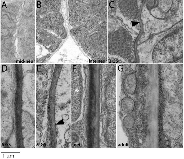
Figure 12.

Development of the perineural sheath. In all panels, the neural tube is to the right. In panels (A-E), myotome is adjacent to (left of) the perineural sheath, and in panels (F-G), sclerotome-derived mesothelium is present between the myotome and perineural sheath (see text). The perineural sheath develops at the interface between them. A basal lamina is first apparent in 2 GS larvae (C). The black arrowhead in (E) indicates striated collagen fibers, first apparent at the 9 GS stage, and the white asterisk in (F) marks the nucleus of a sclerotome-derived mesothelial cell. All panels are to the scale shown. Stage abbreviations: neur, neurula; GS, gill slit; met, early metamorphosis; adult, subadult.
